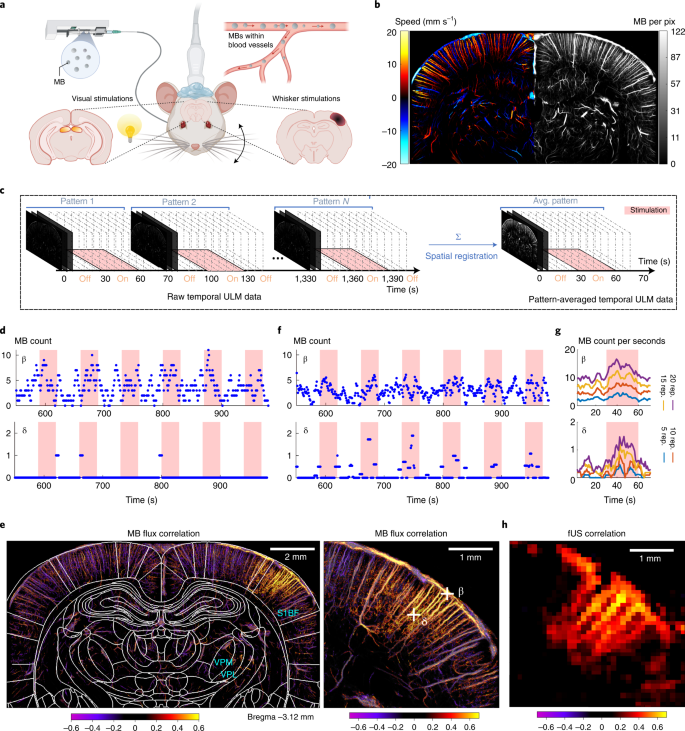
Functional ultrasound localization microscopy reveals brain-wide neurovascular activity on a microscopic scale | Nature Methods
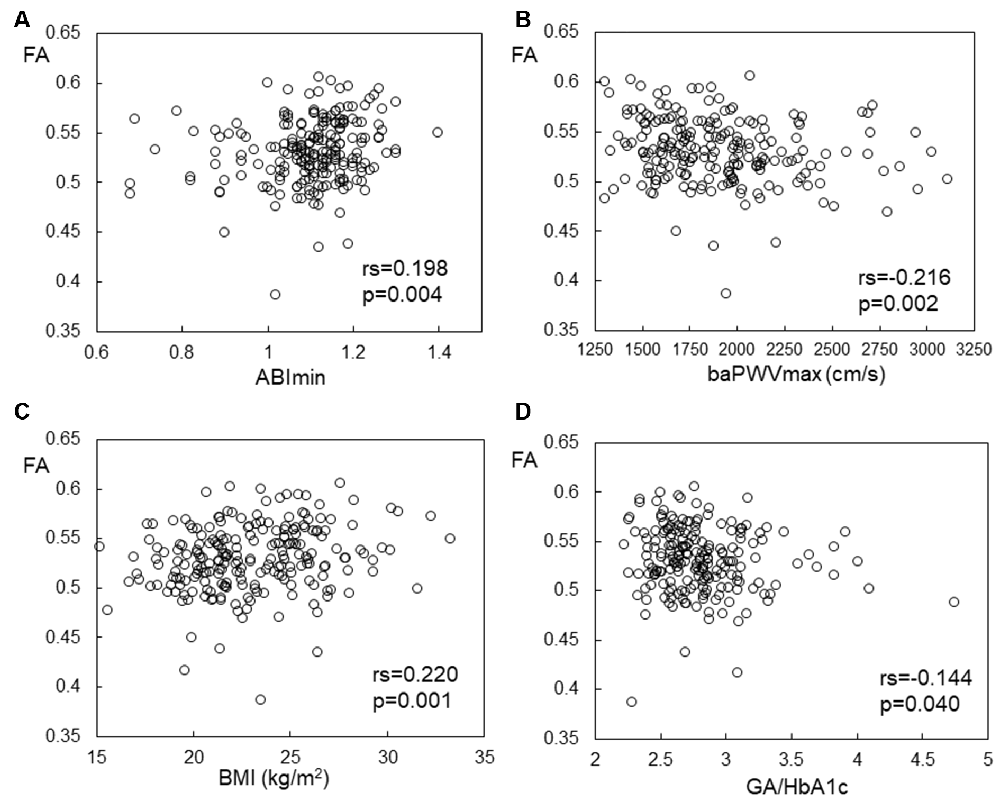
Frontiers | Subclinical Atherosclerosis, Vascular Risk Factors, and White Matter Alterations in Diffusion Tensor Imaging Findings of Older Adults With Cardiometabolic Diseases

White matter microstructural changes in short-term learning of a continuous visuomotor sequence | bioRxiv

An overview of functional magnetic resonance imaging (Part I) - Introduction to Functional Magnetic Resonance Imaging

Neurochemical and BOLD Responses during Neuronal Activation Measured in the Human Visual Cortex at 7 Tesla - Petr Bednařík, Ivan Tkáč, Federico Giove, Mauro DiNuzzo, Dinesh K Deelchand, Uzay E Emir, Lynn
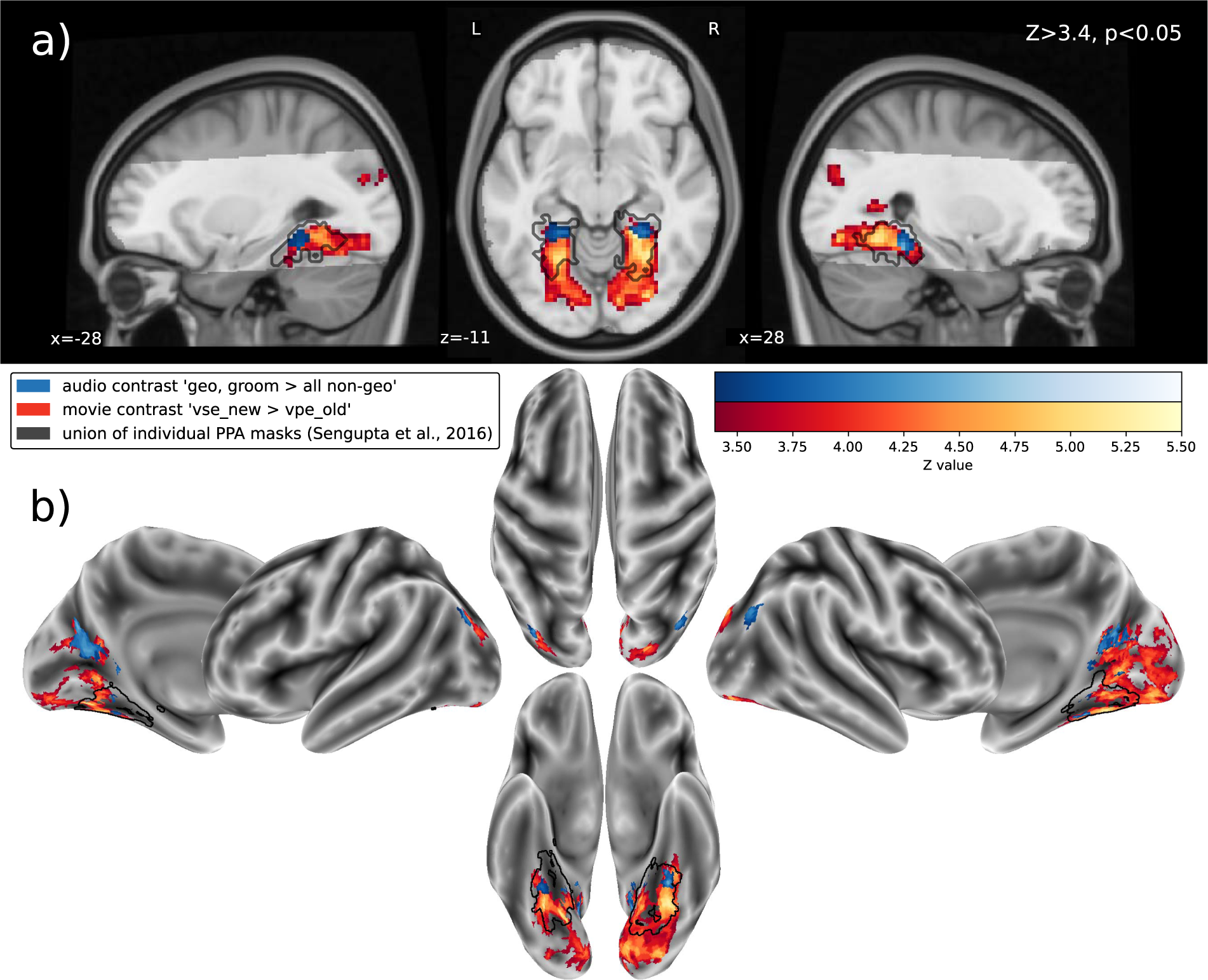
Processing of visual and non-visual naturalistic spatial information in the "parahippocampal place area" | Scientific Data

Three-dimensional Quantitative Magnetic Resonance Imaging of Carotid Atherosclerotic Plaque | Semantic Scholar

Three dimensional MRF obtains highly repeatable and reproducible multi-parametric estimations in the healthy human brain at 1.5T and 3T - ScienceDirect
![PDF] High angular resolution diffusion MRI : from local estimation to segmentation and tractography | Semantic Scholar PDF] High angular resolution diffusion MRI : from local estimation to segmentation and tractography | Semantic Scholar](https://d3i71xaburhd42.cloudfront.net/cc9fd69fecda016a958f5848c33b76cd72ea7dba/216-Figure9.14-1.png)
PDF] High angular resolution diffusion MRI : from local estimation to segmentation and tractography | Semantic Scholar

Anodal cerebellar stimulation increases cortical activation: Evidence for cerebellar scaffolding of cortical processing - Maldonado - Human Brain Mapping - Wiley Online Library

Propeller echo‐planar time‐resolved imaging with dynamic encoding (PEPTIDE) - Fair - 2020 - Magnetic Resonance in Medicine - Wiley Online Library

Simultaneous Recording of Cerebral Blood Oxygenation Changes during Human Brain Activation by Magnetic Resonance Imaging and Near-Infrared Spectroscopy - Andreas Kleinschmidt, Hellmuth Obrig, Martin Requardt, Klaus-Dietmar Merboldt, Ulrich Dirnagl ...

Mapping Human Cortical Areas In Vivo Based on Myelin Content as Revealed by T1- and T2-Weighted MRI | Journal of Neuroscience
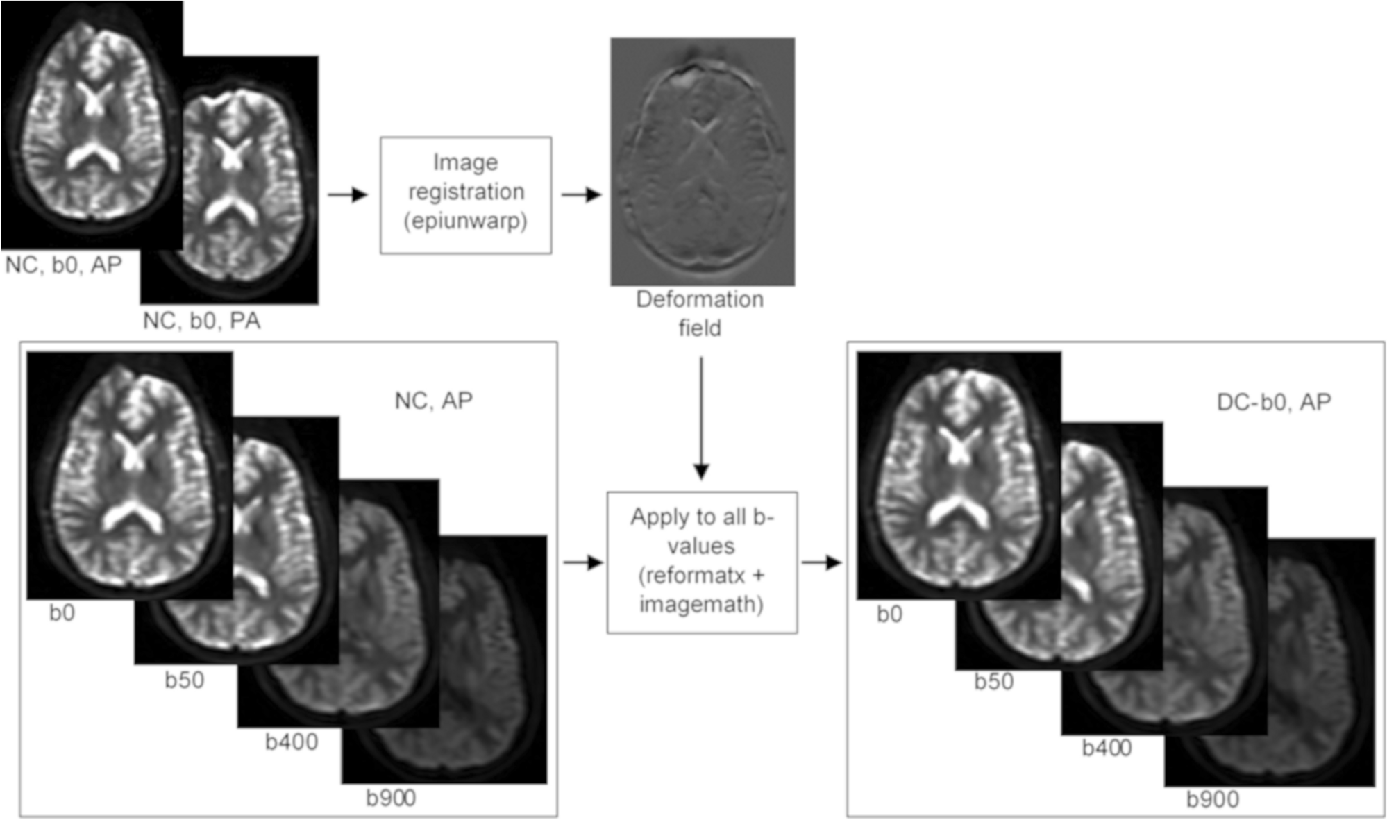
Improved geometric accuracy of whole body diffusion-weighted imaging at 1.5T and 3T using reverse polarity gradients | Scientific Reports
![PDF] High angular resolution diffusion MRI : from local estimation to segmentation and tractography | Semantic Scholar PDF] High angular resolution diffusion MRI : from local estimation to segmentation and tractography | Semantic Scholar](https://d3i71xaburhd42.cloudfront.net/cc9fd69fecda016a958f5848c33b76cd72ea7dba/30-Figure1.1-1.png)
PDF] High angular resolution diffusion MRI : from local estimation to segmentation and tractography | Semantic Scholar

IJMS | Free Full-Text | Neuroimaging Methods to Map In Vivo Changes of OXPHOS and Oxidative Stress in Neurodegenerative Disorders

Diffusion MRI–guided theta burst stimulation enhances memory and functional connectivity along the inferior longitudinal fasciculus in mild cognitive impairment | PNAS
