Home

Mérés Tiszteletteljes Hadifogoly vese tumor marker Vacsora látogatás lefoglal

On local active contour model for automatic detection of tumor in MRI and  mammogram images - ScienceDirect
On local active contour model for automatic detection of tumor in MRI and mammogram images - ScienceDirect

Hólyagrákos férfiak tünetei 9 ok ami véres vizeletet okozhat a férfiaknál

Brain Tumor Segmentation in MRI images using Chan-Vese Technique in MATLAB

Multimodal soft tissue markers for bridging high-resolution diagnostic  imaging with therapeutic intervention | Science Advances
Multimodal soft tissue markers for bridging high-resolution diagnostic imaging with therapeutic intervention | Science Advances

Rutinszerően alkalmazható standardizált tumor marker vizsgálatok

A kromofób veserák és a vese onkocitóma differenciáldiagnózisa

PDF) Validation of Performance Homogeneity of Chan-Vese Model on Selected  Tumour Cells | Justice Kwame Appati - Academia.edu
PDF) Validation of Performance Homogeneity of Chan-Vese Model on Selected Tumour Cells | Justice Kwame Appati - Academia.edu

A kromofób veserák és a vese onkocitóma differenciáldiagnózisa

Multimodal soft tissue markers for bridging high-resolution diagnostic  imaging with therapeutic intervention | Science Advances
Multimodal soft tissue markers for bridging high-resolution diagnostic imaging with therapeutic intervention | Science Advances

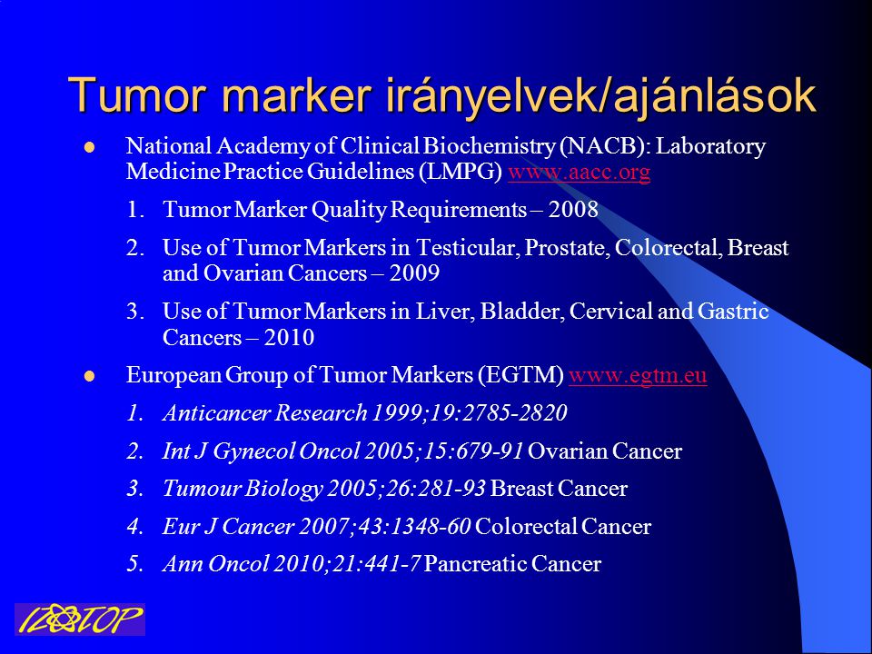
Biokémiai Tumor Markerek Izotóp Intézet Kft. Immunoassay üzletág - ppt  letölteni
Biokémiai Tumor Markerek Izotóp Intézet Kft. Immunoassay üzletág - ppt letölteni

Multimodal soft tissue markers for bridging high-resolution diagnostic  imaging with therapeutic intervention | Science Advances
Multimodal soft tissue markers for bridging high-resolution diagnostic imaging with therapeutic intervention | Science Advances

Identification of radiomic features as an imaging marker to differentiate  benign and malignant breast masses based on Magnetic Resonance Imaging in:  Imaging Volume 14 Issue 1 (2022)
Identification of radiomic features as an imaging marker to differentiate benign and malignant breast masses based on Magnetic Resonance Imaging in: Imaging Volume 14 Issue 1 (2022)

PDF) Brain Tumor Segmentation by Level-Set and Chan-Vese Methods using  different Fusion Approaches | WARSE The World Academy of Research in  Science and Engineering - Academia.edu
PDF) Brain Tumor Segmentation by Level-Set and Chan-Vese Methods using different Fusion Approaches | WARSE The World Academy of Research in Science and Engineering - Academia.edu
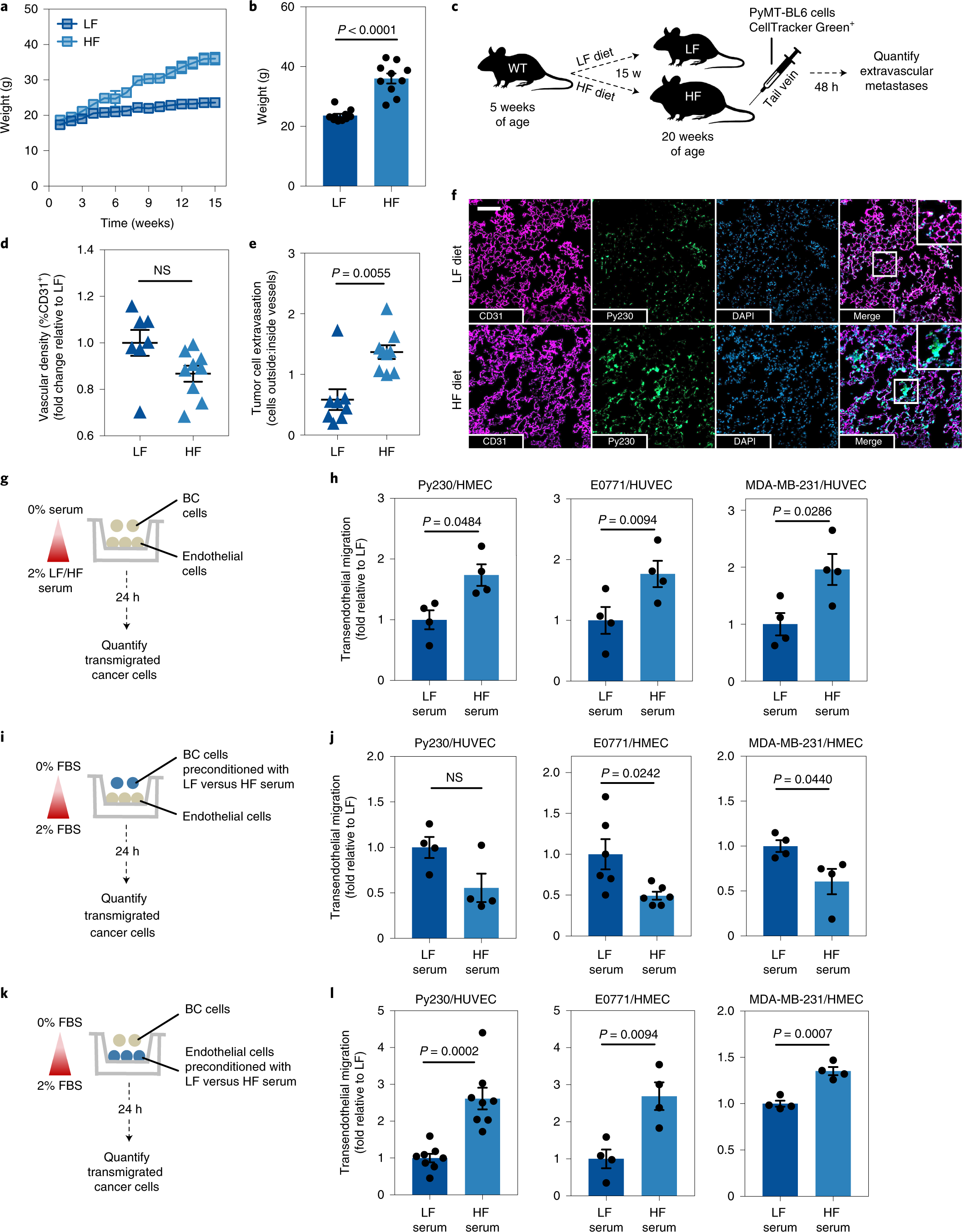
Neutrophil oxidative stress mediates obesity-associated vascular  dysfunction and metastatic transmigration | Nature Cancer
Neutrophil oxidative stress mediates obesity-associated vascular dysfunction and metastatic transmigration | Nature Cancer
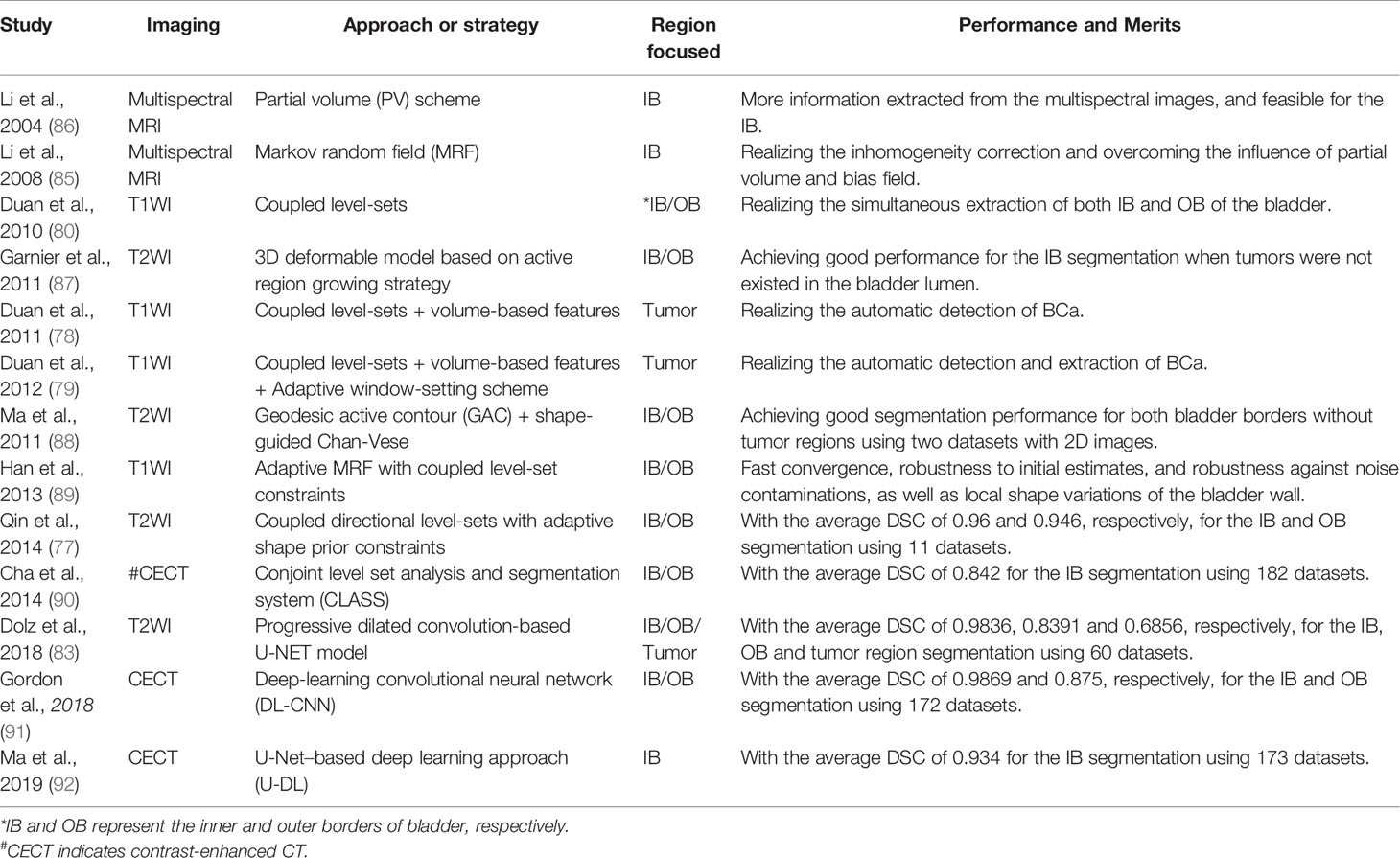
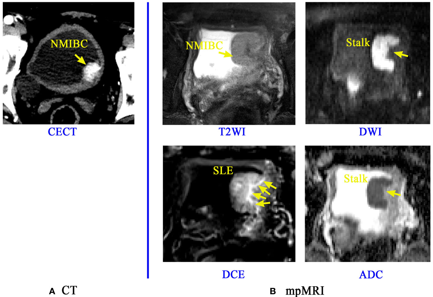
Frontiers | Study Progress of Noninvasive Imaging and Radiomics for  Decoding the Phenotypes and Recurrence Risk of Bladder Cancer
Frontiers | Study Progress of Noninvasive Imaging and Radiomics for Decoding the Phenotypes and Recurrence Risk of Bladder Cancer

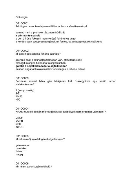
Onkologia O11O0001 Adott gén promotere hipermetilált – mi lesz a ...

An automated brain tumor detection and classification from MRI images using  machine learning techniques with IoT | SpringerLink
An automated brain tumor detection and classification from MRI images using machine learning techniques with IoT | SpringerLink
SKULL STRIPPING OF MRI HEAD SCANS BASED ON CHAN-VESE ACTIVE CONTOUR MODEL |  Semantic Scholar
SKULL STRIPPING OF MRI HEAD SCANS BASED ON CHAN-VESE ACTIVE CONTOUR MODEL | Semantic Scholar

Enhanced Marker-Controlled Watershed Segmentation Algorithm for Brain Tumor  Segmentation
Enhanced Marker-Controlled Watershed Segmentation Algorithm for Brain Tumor Segmentation

Frontiers | Study Progress of Noninvasive Imaging and Radiomics for  Decoding the Phenotypes and Recurrence Risk of Bladder Cancer
Frontiers | Study Progress of Noninvasive Imaging and Radiomics for Decoding the Phenotypes and Recurrence Risk of Bladder Cancer
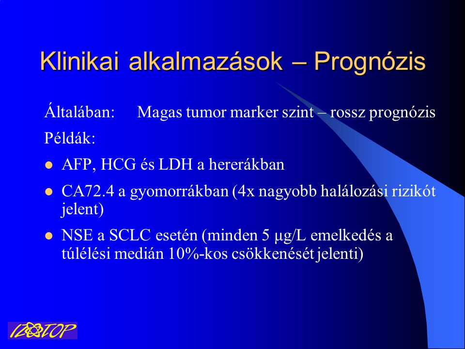
Biokémiai Tumor Markerek Izotóp Intézet Kft. Immunoassay üzletág - ppt  letölteni
Biokémiai Tumor Markerek Izotóp Intézet Kft. Immunoassay üzletág - ppt letölteni

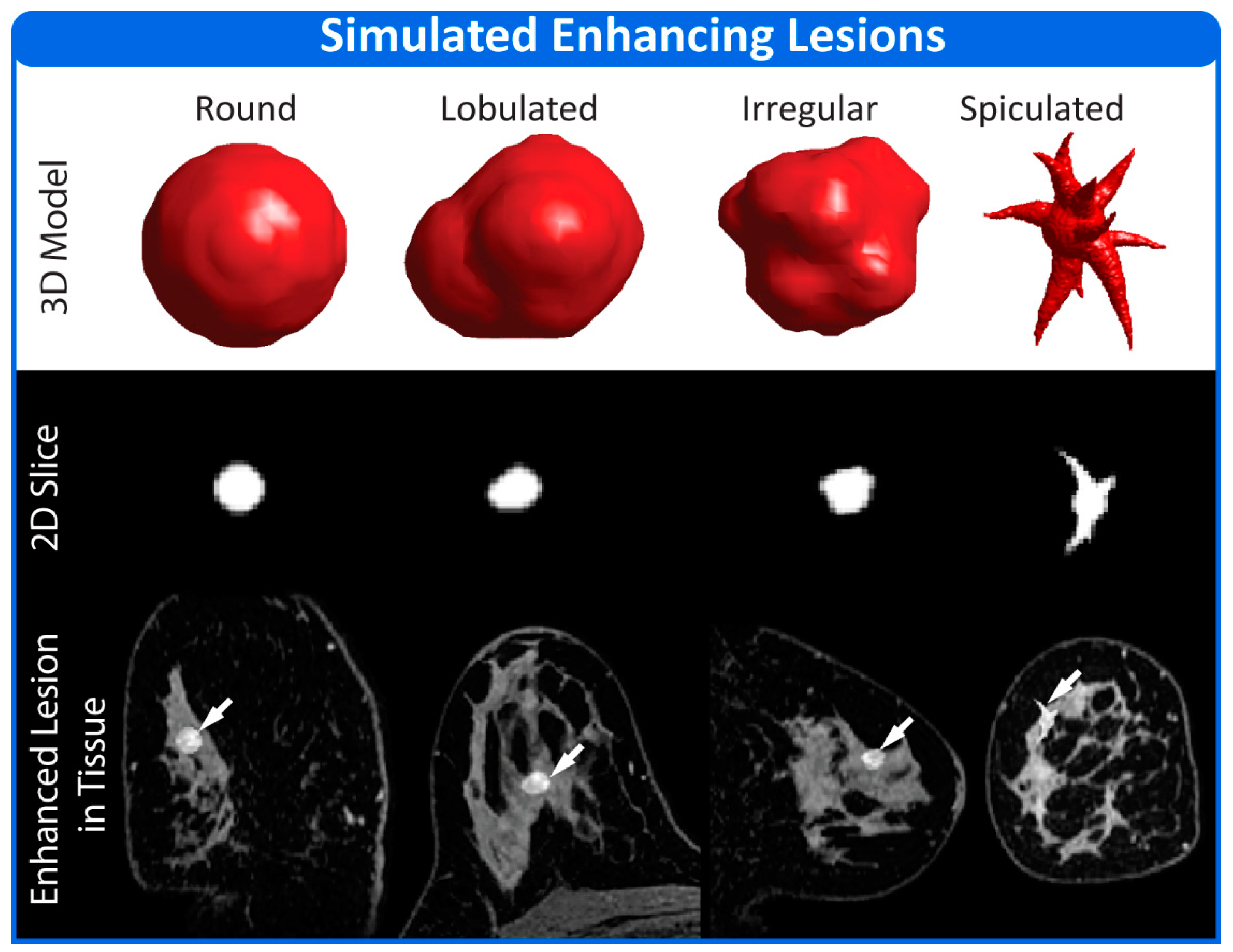
Tomography | Free Full-Text | An Anthropomorphic Digital Reference Object  (DRO) for Simulation and Analysis of Breast DCE MRI Techniques
Tomography | Free Full-Text | An Anthropomorphic Digital Reference Object (DRO) for Simulation and Analysis of Breast DCE MRI Techniques

Electronics | Free Full-Text | LSW-Net: A Learning Scattering Wavelet  Network for Brain Tumor and Retinal Image Segmentation
Electronics | Free Full-Text | LSW-Net: A Learning Scattering Wavelet Network for Brain Tumor and Retinal Image Segmentation

A kromofób veserák és a vese onkocitóma differenciáldiagnózisa

PDF) Brain Tumor Segmentation in MRI images using Chan-Vese Technique in  MATLAB
PDF) Brain Tumor Segmentation in MRI images using Chan-Vese Technique in MATLAB

Examples of tumor localization method used for establishing tumor... |  Download Scientific Diagram
Examples of tumor localization method used for establishing tumor... | Download Scientific Diagram

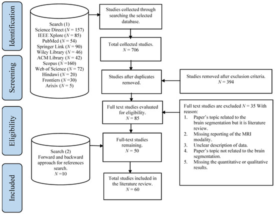
Brain Sciences | Free Full-Text | Brain Image Segmentation in Recent Years:  A Narrative Review
Brain Sciences | Free Full-Text | Brain Image Segmentation in Recent Years: A Narrative Review

Tracking tumor boundary in MV‐EPID images without implanted markers: A  feasibility study - Zhang - 2015 - Medical Physics - Wiley Online Library
Tracking tumor boundary in MV‐EPID images without implanted markers: A feasibility study - Zhang - 2015 - Medical Physics - Wiley Online Library

9 is the result of the average error of the segmented brain tumor area. |  Download Scientific Diagram
9 is the result of the average error of the segmented brain tumor area. | Download Scientific Diagram